Technology

Clinical Programs

Explore clinical use cases, evidence milestones, and collaboration pathways for early adopters and trial partners.

A dramatic cinematic close-up of a transparent, holographic whole-body bioimpedance tissue map floating above a sleek black glass table in a dimly lit research lab. The semi-transparent body silhouette is rendered in luminous gradients of cyan, teal, and amber, with distinct organs and tissues outlined as functional conductivity patterns rather than anatomy slices. Around the hologram, thin filaments of light represent AI networks performing real-time reconstruction, connecting to a minimal, unbranded silver workstation. Narrow beams of cool white light from above catch faint mist in the air, while the background fades into a soft bokeh of lab equipment and dark cabinetry. Framed using the rule of thirds with a shallow depth of field, the mood is analytical yet visionary, highlighting radiation-free, multimodal functional mapping with cinematic realism.
A sleek, futuristic immersion bioimpedance imaging chamber occupies the center of a dark, high-tech clinical suite, its transparent cylindrical tank filled with crystal-clear water faintly tinged aqua. Within the chamber, a minimalist, non-human mannequin form made of matte white composite material is suspended weightlessly, ringed by slender metallic electrode bands and subtle sensor arrays. Around the room, large wall-mounted displays show glowing whole-body functional tissue maps in rich blues, teals, and ambers, emphasizing radiation-free imaging. Cinematic, cool-toned overhead lighting and soft floor uplights carve crisp reflections on polished black surfaces, creating a precise, confident atmosphere. Shot at eye level with a wide-angle lens, the composition emphasizes depth and symmetry, with photographic realism and a clean, cinematic aesthetic that communicates cutting-edge, AI-powered precision medicine.

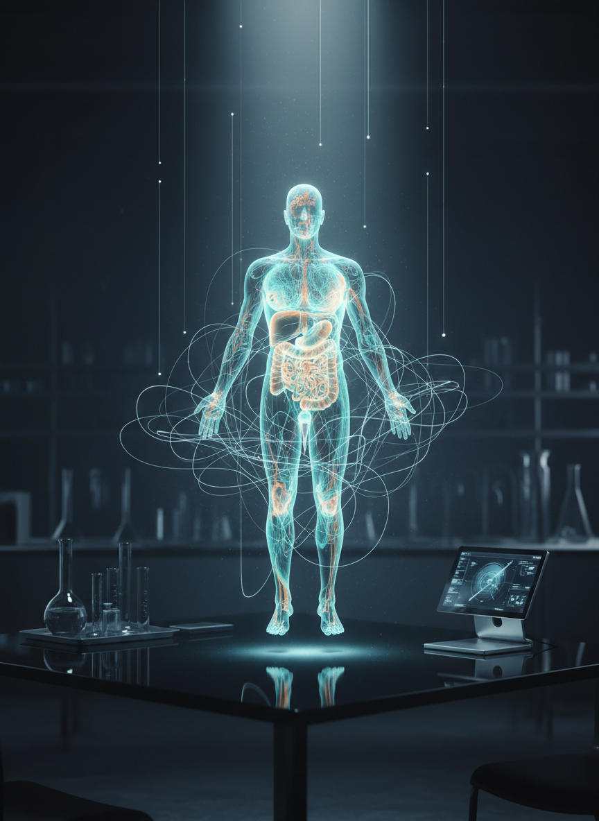
Radiation-Free Clinical Applications

Whole-body bioimpedance imaging supports oncology staging, treatment response monitoring, organ function assessment, and perioperative risk stratification with radiation-free, repeatable scans, designed for integration into hospital workflows and prospective trials across radiology, oncology, and critical care. See resources for protocols and data.

Solutions

An expansive, cinematic bird’s-eye view of a precision-medicine command center, dominated by a curved, wall-to-wall display showing synchronized panels of whole-body bioimpedance maps, MRI outlines, and ultrasound silhouettes seamlessly fused. Each panel glows with scientifically accurate, color-coded functional tissue maps in cool blues and warm golds, conveying subtle physiological differences rather than anatomical cuts. In the center foreground, a single unbranded, matte-white central console with gesture controls faces the display, hinting at AI-guided decision support without showing any humans. Overhead spotlights cast soft pools of light onto a matte graphite floor, while the rest of the room remains in shadow, evoking focus and discretion. Shot with a wide cinematic lens, the composition emphasizes scale and integration, ideal for an investor-focused overview of AquaBIT AI’s multimodal platform.

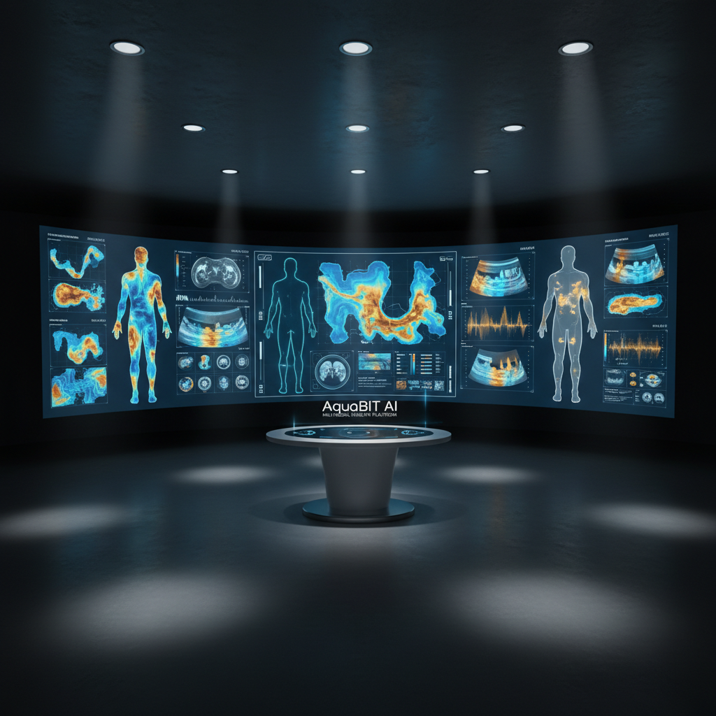
Oncology decision-support workflow integrating AquaBIT imaging, reporting templates, and multidisciplinary review for staging, therapy selection, and longitudinal surveillance.

A dramatic cinematic close-up of a transparent, holographic whole-body bioimpedance tissue map floating above a sleek black glass table in a dimly lit research lab. The semi-transparent body silhouette is rendered in luminous gradients of cyan, teal, and amber, with distinct organs and tissues outlined as functional conductivity patterns rather than anatomy slices. Around the hologram, thin filaments of light represent AI networks performing real-time reconstruction, connecting to a minimal, unbranded silver workstation. Narrow beams of cool white light from above catch faint mist in the air, while the background fades into a soft bokeh of lab equipment and dark cabinetry. Framed using the rule of thirds with a shallow depth of field, the mood is analytical yet visionary, highlighting radiation-free, multimodal functional mapping with cinematic realism.

Functional organ assessment package combining acquisition protocols, AI analytics, and EHR integration for cardiology, nephrology, and metabolic disease programs.

Upcoming Studies

2025-10-05

Phase I Center

Boston, MA

2025-10-20

Oncology Congress

Chicago, IL

2025-11-08

AI Imaging

Paris, France

2025-11-12

Investigator Summit

Toronto, ON

2025-11-03

Clinical Trials

San Diego, CA

2025-12-02

KOL Roundtable

Zurich, Switzerland

2026-02-18

Imaging Workshop

London, UK

2026-01-27

Radiology Symposium

Berlin, Germany

2026-03-10

Translational Center

Singapore General