Investigational · Oncology first
Investigating cancer through its electrical properties
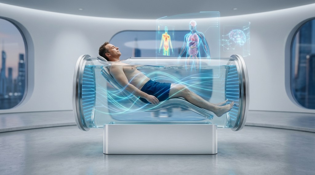
AquaBIT is developing water-assisted, multi-frequency bioimpedance imaging and physics-informed AI for repeatable functional tissue mapping in oncology research.
Current stage: computational feasibility, tissue-equivalent phantoms and prototype engineering. AquaBIT is not yet a clinically validated or approved medical device.
INTERACTIVE OVERVIEW
From electrical signal to longitudinal oncology insight
Explore the proposed AquaBIT pathway. Each stage links to the evidence, technology and clinical questions that must be addressed.
AquaBIT is investigational and is not approved for diagnosis, treatment selection or patient management.
The clinical challenge
Cancer treatment increasingly needs measurements over time
CT, MRI, PET, ultrasound and histopathology remain indispensable. Yet longitudinal oncology care can require repeated assessments, and every modality brings different constraints in radiation, tracers, contrast, capacity, cost or anatomical coverage.
AquaBIT asks whether tissue electrical properties can provide an additional, repeatable source of functional information—complementing established imaging rather than competing with it.
Why AquaBIT is different
Water-assisted acquisition
Water creates a continuous conductive interface around the body, potentially reducing the contact variability associated with multiple skin electrodes and enabling distributed measurements from many directions.
Multi-frequency measurement
Electrical responses are measured across frequencies because conductivity, permittivity, membrane behaviour and fluid distribution may contribute different information to the acquired signal.
Physics-informed reconstruction
Finite-element models, regularised inverse methods and AI are being combined to reconstruct candidate conductivity maps while preserving calibration, uncertainty and physical consistency.
Oncology first: treatment-response research
The initial clinical proposition is deliberately focused: investigate whether reproducible changes in electrical tissue properties can contribute to monitoring response during systemic anticancer therapy.
Repeatable
No ionising radiation is generated by the AquaBIT measurement itself, supporting investigation of more frequent serial assessment.
Quantitative
The research output is intended to include traceable regional measurements, longitudinal comparison and explicit uncertainty—not merely a visual image.
Complementary
Candidate biomarkers would be evaluated against MRI, CT, PET, ultrasound, pathology and clinical outcomes as appropriate to the tumour and study.
A staged route to evidence
01
Model
Simulate current propagation, tumour contrast and electrode sensitivity.
02
Measure
Recover known targets in calibrated tissue-equivalent phantoms.
03
Integrate
Build the chamber, electronics, safety controls and reconstruction pipeline.
04
Validate
Compare reproducibility and clinical signal with appropriate reference standards.
Clinical leadership, multidisciplinary development
Dr Juan Martin
Founder and Clinical Lead
Intensive Care Consultant and clinical innovator leading the medical vision, safety principles and translational strategy for AquaBIT.
Tahmid Rudman
Project Management
Supporting programme coordination, development planning and the multidisciplinary pathway from concept through technical and clinical feasibility.
Specialist collaborators
Building the evidence team
AquaBIT is engaging expertise across oncology, medical physics, computational imaging, biomedical engineering and clinically aligned AI.
Help establish what AquaBIT can—and cannot—measure
We welcome rigorous collaboration with oncology centres, universities, engineers, AI researchers, medical-device partners and early-stage healthcare investors.

