ONCOLOGY RESEARCH PROGRAMME
From electrical measurements to clinically meaningful evidence
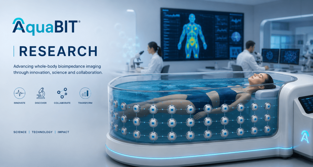
AquaBIT is investigating whether water-assisted, multi-frequency bioimpedance measurements can support repeatable functional tissue mapping and treatment-response research in solid tumours.
A disciplined translational pathway
The central question is not simply whether tissues differ electrically. It is whether those differences can be measured reproducibly, reconstructed reliably and linked to outcomes that matter in oncology.
AquaBIT therefore follows a staged programme: computational feasibility, controlled phantom validation, prototype engineering and prospective clinical research. Each stage must meet predefined technical and safety criteria before the next begins.
AquaBIT remains investigational. It is not approved for diagnosis, treatment selection or patient management.
RESEARCH ROADMAP
Four stages, one evidence standard
01. Model
Physics-based human-body and tumour models test signal detectability, electrode geometry, frequency selection and reconstruction assumptions before hardware studies.
02. Validate
Tissue-mimicking phantoms and controlled experiments assess sensitivity, repeatability, calibration drift and the effect of motion and biological variability.
03. Engineer
The water-assisted electrode array, multi-frequency electronics and quality-control pipeline are integrated into a safe, reproducible research prototype.
04. Study
Ethics-approved feasibility studies will assess safety, usability, signal quality and agreement with reference imaging before larger multicentre evaluation.

COMPUTATION + ENGINEERING
Reconstruction must be explainable and testable
Bioimpedance data are indirect and sensitive to geometry, contact conditions and modelling assumptions. Our programme combines finite-element modelling, physics-informed reconstruction and machine learning with explicit quality controls.
- Multi-frequency signal acquisition and calibration
- Forward-model and reconstruction error analysis
- Repeatability and uncertainty reporting
- Comparison with MRI, CT, ultrasound or pathology where appropriate
- Prospective validation on data not used for model development
INITIAL ONCOLOGY PRIORITY
Repeatable treatment-response research
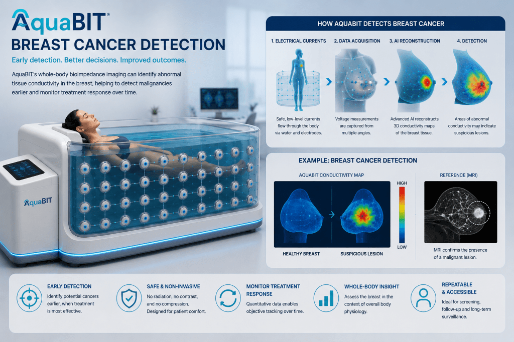
The first clinical hypothesis is that serial conductivity maps may reveal functional changes during systemic therapy or radiotherapy. The aim is to investigate a complementary research signal—not to replace established imaging or pathology.
- Breast and other accessible solid tumours
- Within-patient longitudinal measurement
- Correlation with established response criteria
- Early assessment of reproducibility and clinical workflow
What success must demonstrate
Technical
Stable acquisition, measurable contrast, reconstruction accuracy, repeatability and transparent uncertainty.
Clinical
Safe operation, acceptable workflow, interpretable outputs and meaningful comparison with reference standards.
Translational
Prospective evidence, multicentre reproducibility, regulatory readiness and a defined clinical-use pathway.
Scientific foundations and selected primary evidence
Published research establishes that biological tissues have frequency-dependent dielectric properties, that breast-tissue measurements vary between normal and pathological samples, and that three-dimensional electrical impedance tomography can reconstruct conductivity distributions. These studies support the scientific basis for measurement and reconstruction research; they do not establish the clinical performance of AquaBIT.
- Gabriel C, Gabriel S, Corthout E. The dielectric properties of biological tissues: I. Literature survey (1996)
- Jossinet J. Variability of impedivity in normal and pathological breast tissue (1996)
- Jossinet J. The impedivity of freshly excised human breast tissue (1998)
- Cherepenin VA et al. A 3D electrical impedance tomography system for breast cancer detection (2001)
- Halter RJ et al. In vivo and ex vivo tissue dielectric properties for breast imaging (2009)
- Boverman G et al. Multifrequency EIT reconstruction of the breast (2008)
Build the evidence with us
We welcome serious discussions with oncology centres, biomedical engineers, computational-imaging groups, trial methodologists and strategic research partners.
Review the evidence library
See how AquaBIT distinguishes concept illustrations, simulations, phantom work and future measured results.
Solve an evidence question with us
Defined collaboration pathways are available for oncology centres, engineers and computational-imaging teams.
The research rationale in one document
The AquaBIT White Paper sets out the biophysical basis, testable oncology hypotheses, measurement pipeline, limitations and staged validation programme.
Human Electrical–Molecular Atlas
Every paired dataset strengthens the research platform.
AquaBIT’s long-term research programme will connect electrical tissue behaviour with blood-derived molecular profiles, reference imaging, diagnosis and outcomes. This governed atlas could support digital-biomarker discovery, longitudinal comparison and future biological foundation models.
Explore the Human Atlas programme
This is an AquaBIT research ambition and does not imply current clinical deployment or institutional endorsement.

