Initial clinical research focus
A focused first proposition: measuring treatment response over time
AquaBIT is investigating whether serial changes in electrical tissue properties can provide complementary functional information during systemic anticancer therapy—without adding ionising radiation from the AquaBIT measurement itself.
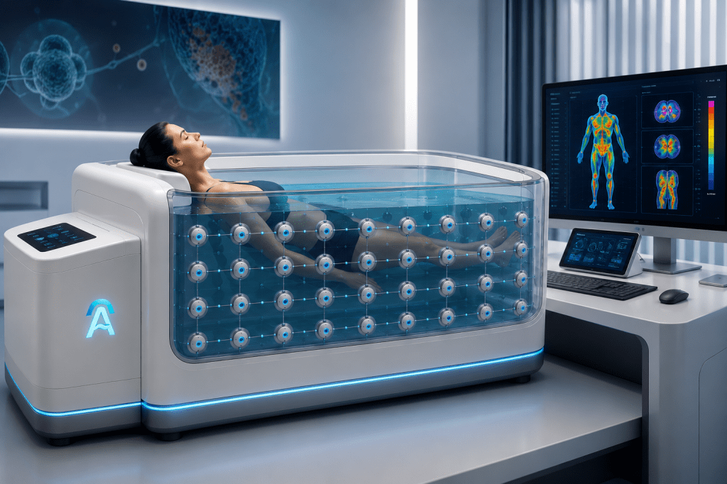
Development-stage statement: AquaBIT is an investigational research platform. It does not currently detect, diagnose, stage or monitor cancer in patients, and it is not approved to guide treatment.
Why treatment-response monitoring?
Oncological response is not a single event. Tumours and normal tissues can evolve during chemotherapy, immunotherapy, targeted treatment and radiotherapy, while structural size changes may not capture every relevant biological effect.
The initial AquaBIT hypothesis is that frequency-dependent electrical changes may contain information related to tissue water, cellularity, membrane integrity, necrosis, vascularity or extracellular structure. Whether those signals are detectable and clinically useful must be established experimentally.
The proposed clinical role
Baseline characterisation
Acquire a reproducible pre-treatment electrical-property dataset for comparison with later examinations.
Serial measurement
Investigate whether regional or whole-body signals change during therapy and whether the direction and magnitude are reproducible.
Reference comparison
Compare candidate AquaBIT biomarkers with established imaging, pathology, laboratory markers and clinical outcomes.
SAME-VISIT MONITORING CONCEPT
Every chemotherapy visit could become a treatment-response checkpoint
Patients already return to the oncology unit for successive chemotherapy cycles. Subject to clinical validation, AquaBIT could be incorporated into those scheduled attendances, creating a patient-specific series of measurements rather than waiting for widely spaced conventional imaging appointments.
01 · BEFORE THERAPY
Establish baseline
A standardised pre-treatment study creates the patient-specific reference against which later changes may be evaluated.
02 · EACH ATTENDANCE
Repeat consistently
A brief acquisition could be scheduled at each chemotherapy visit, using controlled timing, positioning, hydration and measurement conditions.
03 · RAPID COMPARISON
Measure the trend
Quality-controlled reconstruction could compare the new dataset with baseline and previous cycles, highlighting the direction and magnitude of candidate changes.
04 · CLINICAL CONTEXT
Inform the next step
A same-visit research report could be reviewed alongside symptoms, examination, laboratory markers and established imaging—not used in isolation.
The potential: a near-real-time signal of response
If serial electrical-property changes are proven to correlate with tumour response, clinicians could receive an earlier indication that treatment appears effective—or that confirmatory assessment may be needed—while the patient is still moving through the chemotherapy pathway. This could shorten the delay between biological change and clinical recognition.
This remains a research hypothesis. AquaBIT cannot currently determine whether chemotherapy is working. Prospective studies must establish repeatability, clinically meaningful thresholds, timing effects and agreement with accepted response criteria before this information could support patient management.
Questions the research must answer
- What conductivity or impedance contrast is realistically detectable?
- What spatial resolution can be achieved at clinically relevant depths?
- How stable are measurements across position, temperature and time?
- Can tumour-related signals be separated from normal physiological variation?
- Do electrical changes correlate with pathological or radiological response?
- Does AquaBIT add clinically useful information beyond existing pathways?
A disciplined validation pathway
- In silico: computational models and sensitivity analysis across candidate tumour sites.
- Phantom: blinded recovery of known targets, contrasts, depths and volumes.
- Technical feasibility: repeatability, calibration, safety and human-factors evaluation.
- Clinical feasibility: prospective acquisition alongside standard-of-care investigations.
- Clinical validation: predefined performance thresholds and multicentre external validation.
Partner with the oncology research programme
We welcome collaboration on tumour modelling, phantom design, imaging biomarkers, study methodology, clinical pathways and prospective validation.
Future oncology roadmap
From molecular signal to spatial investigation.
Blood-derived cfDNA, methylation, protein or metabolic patterns could be studied beside AquaBIT’s longitudinal electrical phenotype. AI may associate the measured domains and guide definitive investigation with MRI, CT, PET, ultrasound or pathology where appropriate.
Explore the Molecular oncology pathway
Future research concept. AquaBIT does not directly measure DNA, proteins or metabolites and is not a replacement for pathology or established imaging.

