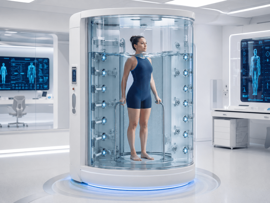
AquaBIT is being developed as an integrated medical-imaging architecture that connects signal acquisition, computational modelling, artificial intelligence and clinical information systems within a single platform. During each examination, a high-density array of electrodes positioned around the water-filled chamber delivers controlled, low-amplitude alternating currents and records the resulting voltage responses across multiple frequencies. Water provides a stable conductive interface around the body, while the acquisition system captures large numbers of spatially distributed measurements that reflect differences in tissue conductivity, permittivity and frequency-dependent impedance. These raw signals form the primary dataset from which AquaBIT reconstructs volumetric representations of internal tissue properties.
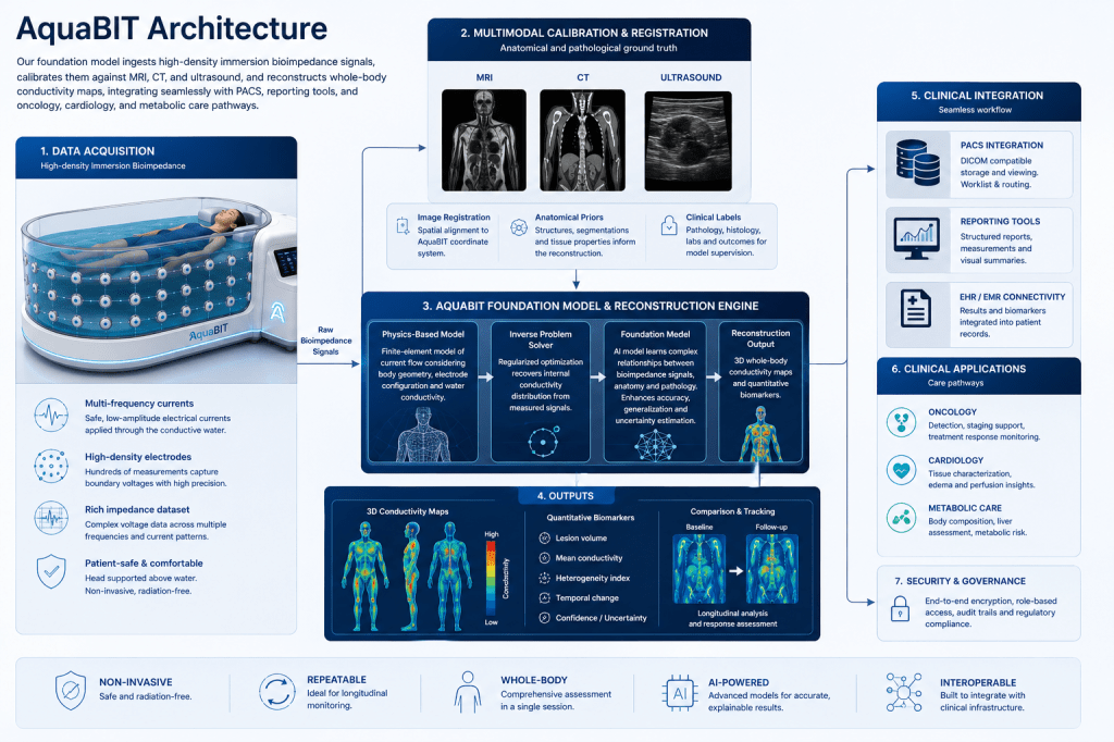
The reconstruction engine combines a physics-based model of the chamber, electrode geometry, water conductivity and human anatomy with advanced inverse-problem algorithms. Finite-element modelling is used to estimate how electrical current propagates through the body, while regularisation methods reduce instability and measurement noise during image reconstruction. A foundation model then learns relationships between bioimpedance measurements, anatomical structures and pathological tissue patterns. Rather than relying exclusively on data-driven prediction, the proposed architecture incorporates physical constraints, calibration parameters and uncertainty estimates so that reconstructed conductivity maps remain consistent with both the acquired electrical signals and established principles of bioelectricity.
MRI, CT and ultrasound data provide anatomical reference information during development and validation. These modalities may be spatially registered with AquaBIT datasets to create paired multimodal training data, allowing the model to learn how electrical-property distributions correspond to recognised anatomical structures and clinically characterised abnormalities. Histopathology, laboratory findings and longitudinal clinical outcomes could provide additional reference standards for disease-specific applications. Once sufficiently validated, the model may reconstruct AquaBIT images directly from immersion-bioimpedance data while using prior multimodal information to improve anatomical localisation, tissue classification and confidence estimation. This process is intended to produce quantitative three-dimensional conductivity maps, regional biomarkers and comparison tools for serial examinations.
The clinical architecture is designed to integrate with existing hospital workflows rather than operate as an isolated diagnostic system. Reconstructed images and structured results could be transmitted to picture archiving and communication systems using standard medical-imaging formats, while quantitative findings could populate reporting software and electronic patient records. Application-specific analytical modules could support oncology through lesion characterisation and treatment-response monitoring, cardiology through the assessment of fluid distribution and tissue properties, and metabolic medicine through longitudinal analysis of body composition. The ultimate objective is to create a modular and interoperable platform in which a common whole-body foundation model supports multiple validated clinical solutions without compromising data traceability, cybersecurity, regulatory oversight or clinician interpretation.
Hardware Architectural Models