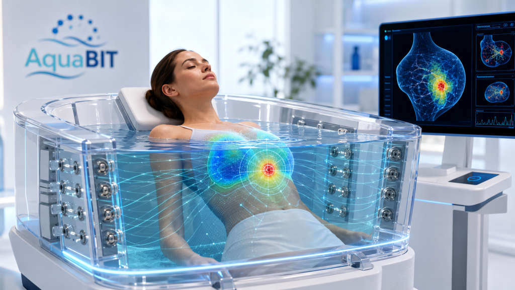
Breast Cancer Detection with AquaBIT
Breast cancer assessment currently depends on mammography, ultrasound, magnetic resonance imaging and biopsy. These methods are clinically established, but each has limitations related to radiation exposure, breast compression, operator dependence, contrast administration, cost or accessibility. AquaBIT is being developed as a complementary imaging platform that investigates whether abnormalities in breast tissue can be identified through their electrical properties. The system is not intended to replace mammography, MRI, ultrasound or histopathology at this stage. Its scientific value would need to be demonstrated through rigorous technical validation and prospective clinical studies.
The scientific basis of AquaBIT is that biological tissues have measurable, frequency-dependent electrical characteristics. Conductivity, permittivity, resistance, reactance and phase response are influenced by tissue water content, cellular density, membrane integrity, vascularity and extracellular structure. Malignant breast tissue may therefore differ electrically from surrounding adipose or fibroglandular tissue. AquaBIT proposes to measure these differences by delivering very low-amplitude alternating currents through a controlled water environment and recording the resulting voltage changes with a surrounding electrode array. The acquired data would then be reconstructed into three-dimensional maps of electrical conductivity and tissue heterogeneity using finite-element modelling, regularised inverse reconstruction and artificial intelligence.
In breast cancer research, AquaBIT could be explored for several potential applications. It may help identify focal regions of electrical asymmetry that warrant further assessment with standard imaging, support serial monitoring during neoadjuvant chemotherapy or targeted therapy, and investigate whether changes in conductivity or impedance spectra precede measurable reductions in tumour size. It could also provide complementary information in women with dense breasts, where mammographic sensitivity may be reduced, and contribute to translational research by correlating bioelectrical signatures with tumour grade, receptor status, vascularity, necrosis, stromal composition and pathological response. These possibilities remain investigational and should be framed as research hypotheses rather than established clinical claims.
The proposed development pathway would begin with computational breast models, tissue-equivalent phantoms and ex vivo tissue characterisation before progressing to healthy-volunteer feasibility studies, diagnostic pilot studies and multicentre clinical validation. Key outcomes would include spatial resolution, minimum detectable lesion size, repeatability, sensitivity, specificity, false-positive rates and incremental value alongside mammography, ultrasound and MRI. Patient safety would remain central throughout, with strict control of electrical current, water temperature, infection prevention and assisted transfer. The patient’s head, eyes, nose and mouth would remain outside the water at all times. The long-term objective is to determine whether AquaBIT can generate reproducible, clinically meaningful and radiation-free imaging biomarkers that support more frequent monitoring and more personalised breast cancer care.

